
Highlights
• Autism appears to be a condition that stems from early developmental disturbances in the womb
• Symptoms may be aggravated by exposure to inflammatory substances that are not well tolerated by these individuals
• Autism (ASD) and ADHD (attention-deficit/hyperactivity disorder) tend to co-occur in families and within individuals
• Genetics, exposure to toxins, and maternal autoimmune disease appear to raise the risk of related behavioral conditions
• Autistic behavior is strongly associated with accumulated heavy metals in the brain, likely a result of many different sources combined with certain antioxidant deficiencies
• Metals such as aluminum and mercury require many years or even decades to release from the body
• Suspected environmental toxins include metals found in industrial chemicals, pesticides, infant formula, vaccines, air, soil, and drinking water
• Chelation (elimination of metals from the body/brain) as a potential therapy needs much further research to increase efficacy and prevent disruption of beneficial minerals
• Some cases of autism show recovery over time, though there is no clear pattern of treatment that explains this remediation
• In the future, we may be able to treat autism and other neurological disorders with a variety of nutritional and behavioral therapies optimized for the immune condition of the individual
• We may also be able to predict or prevent developmental disorders by carefully monitoring genetics, environmental exposure, nutrition, and immune health of the mother and fetus before conception and throughout pregnancy
Overview
Autism presents the largest keyhole that we’ve ever found for peeking at the interactions between genetics, environment, and behavior. We may be able to apply the latest research on autism to a wide range of social and learning disabilities that prevent children from realizing their full potential. Some of the same environmental risk factors for autism apply to other conditions such as ADHD, dyslexia, speech and language impediments, motor control and coordination, schizophrenia, chronic fatigue syndrome, autoimmune thyroiditis, multiple sclerosis, Alzheimer’s and Parkinson’s disease, and various cancers. Studies have proposed that overlapping problems with social motivation affect both autism and ADHD, reducing a child’s collaborative activity and increasing reward activity. In fact, the new 2013 diagnosis criteria by the American Psychiatric Association now allow concurrent diagnoses of ADHD and autism. These are very small steps toward a more holistic understanding of disabilities that have far-reaching effects on our society and human progress.
Autism broadly describes a wide range of abnormal social and intellectual behavior that first appears in early childhood and is more prevalent in boys. Autism diagnoses have skyrocketed along with increased awareness and therapeutic resources. Individual susceptibility appears to develop during pregnancy, but symptoms are not recognized until months or years have passed. Many genes and gene mutations are being linked to autism, and the variety of differences in the condition may soon require diverse subtypes and treatments. Furthermore, the underlying imbalances in autism can extend beyond the brain to include epilepsy, inflammatory bowel disease, type 1 diabetes, and mitochondrial dysfunction.
New discoveries reveal how autism associates with a challenged and vulnerable immune system including a leaky gut, reduced antioxidant production and activity, and difficulty releasing heavy metals. Accumulation of metals appears unique to individuals based on genetic susceptibility, immune health, and interaction with other compounds. Risk of autism also increases in the womb with exposure to chlorinated solvents, testosterone, certain drugs, and infectious pathogens. The undeveloped or compromised immune system cannot detoxify these substances. Older fathers increase the risk of related genetic mutations, while mothers with autoimmune disease increase the overall risk of autism.
Metal consumption is largely unrecognized in our diet and environment, as in the case of methylmercury frequently consumed from certain types of fish. Childhood vaccines contained mercury-based preservative thimerosal until 2001, but pervasive aluminum exposure in foods and drugs may pose even greater threats to the brain. Excessive aluminum can be consumed from infant formula, intravenous feeding, vitamin and drug additives, food such as leavened baked foods and processed cheese, aluminum cookware, antacids, deodorant, dental amalgams, certain types of fish, air pollution, and drinking water. Scientists cannot easily study brain tissue of autistic children, but we continue to witness changes between diet and behavior.
Autistic and other challenging behaviors are often influenced by nutrition and environment, but we need larger, long-term studies necessary to evaluate treatments including gluten-free and casein-free diets. New research reveals promising treatment with antioxidants like N-acetyl cysteine, amino acids, essential fatty acids, and probiotics that can help the body detoxify heavy metals and strengthen the gut and immune function. Maternal folic acid consumption may also play a protective role in pregnancy.
Defining autism
We use the term “autism” to label a set of developmental challenges with multiple symptoms including social awkwardness and unresponsiveness, restricted or repetitive behavior, lack of eye contact, and delayed speech. While most symptoms relate to brain function, associated physical conditions can include allergies, seizures, sleep disturbances, and gastrointestinal problems. Autism was first recognized around 1943, and symptoms were initially classified as schizophrenia. The diagnostic criteria for autism continues to change, making disease trends difficult to analyze over time. The average age of diagnosis for autistic spectrum disorders is decreasing to around age 4 or 5, but signs can now be recognized in infancy.
Autistic children have a much higher incidence of abnormal development, reactions to vaccines, allergic and autoimmune disease, and psychiatric conditions. New research proposes that autistic children experience a sort of brain allergy as a result of autoimmune attacks on “escaped” mitochondrial DNA. Autoimmunity is also experienced in the the gut, and autistic patients suffer from more undigested food, bacteria, and other toxic substances leaking out of the intestines and into the blood. A significant number of autistic children have elevated levels of anti-transglutaminase antibodies which are associated with almost all cases of celiac disease, and many autistic cases have improved with gluten-free diets. Research supports the use of food intolerance testing (IgG, not IgE food allergy testing) to identify cases of autism that might benefit from gluten or casein (dairy)-free diets. This is all difficult for parents when their autistic children’s bone mass density and weight can be much lower than average.
The body chemistry of autism displays abnormal levels of hormones that control brain functions, including higher peaks and delayed recovery of cortisol. Interestingly, the cortisol differences are more pronounced in older children with autism when studying cooperative play, underlining the potential of early intervention to improve social behavior. High levels of serotonin are also consistently correlated with autism, and some researchers have explained this by a deficiency of enzyme monoamine oxidase (MAO). This enzyme is controlled by chromosomes, and females naturally have more MAO activity than males. MAO normally degrades or “detoxifies” monoamine neurotransmitters with the help of sulfur and methyl-based amino acids; when there is a deficiency or blocking of MOA, this detoxification activity is inhibited. A proposed risk for MAO deficiency might be excessive early life exposure to substances that require MAO enzymes or their “fuel supply” of amino acids.
In light of the current research, autistic symptoms appear to require both a susceptible fetus and environmental or dietary toxins which damage fragile tissue in certain areas of the brain. A new study found a significant number of autism cases associated with toxic metals in blood and urine, with cadmium and mercury were the most strongly associated. Other metals at issue included lead, thallium, tin, and tungsten. Some of these metals may explain the overlap with other developmental disorders. In fact, researchers found that lead significantly predicted ADHD diagnosis. Manganese was also associated with ADHD behavior.
Incriminating toxins and metals is not so simple, as they form different compounds which are each handled differently by the body based on the immune response. Precise measurement of metal accumulation in live brain tissue – including young children and fetuses – is currently impossible, so scientists rely on blood, urine, and hair to predict with various levels of reliability. It is also impossible to tell if accumulated substances are part of the cause or effect of autism. Risk awareness, and nutritional intervention before and during pregnancy offer the most hope for prevention.
Debating over-diagnosis

While about 1 in every 400 children has diabetes, 1 in every 50 children has autism. 1 in 10 children have ADHD. We have clearly become a society dependent on these labels, using official-stamped diagnoses to receive insurance coverage, special services, and compassion from our community. Autism incidence rates have gone up at a mind-blowing rate of 1,148% between 1987 and 2007, fueling controversy about over-diagnosis. Surely some parents are driving diagnoses, as seen in a California study where autistic cases clustered in areas where parents were more highly educated.
Yet careful research indicates that the rise cannot be completely explained by diagnoses. A close look at yearly cases in California reveals higher odds in each successive group of children, particularly for high-functioning autistic cases. If we are truly more aware and demanding of diagnoses, it would seem that rates of other well-known developmental disabilities would have soared in tandem. On the contrary, ADHD rates only increased 33% between intervals 1997-1999 and 2006-2008 while autism increased 289%. Parent-reported cases of ADHD had no significant increase between 1997 and 2006 in Western states.
In any case, ongoing abnormal child behavior should not be dismissed as an inflated statistic. Even if we have endured developmental disabilities in years past, we have opportunities to improve these moving forward.
Prenatal predispositions
“Emerging evidence supports the idea that prenatal and early postnatal events such as maternal nutrition and drug and chemical exposure are manifested in health consequences later in life.” –Env Health Perspectives, 2013
Protecting the fetus and newborn from controllable health risks is the most important gift to a child. The fetus has an immature immune system that cannot properly manage exposure to toxic substances – including heavy metals and hazardous chemicals ingested, absorbed, or inhaled by the mother. Early diagnosis is equally important, as an autistic child requires early therapy and nutritional support. Fortunately, many cases of autism can now be diagnosed at infancy. Brain abnormalities such as a loss of white matter brain tissue can be detected at birth with ultrasound scanning to determine a risk of autism. After birth, the autistic child may be prone to adverse reactions to vaccines and abnormal development. Younger autistic children appear to have more trouble releasing the body’s heavy metals than others, and additional protection may be necessary for these individuals.
Genetic variations, mutations and older age of father
An estimated 350 to 400 genes may be involved in determining risk of autism. Variations of these genes can affect how autism presents itself, as in the case of the oxytocin receptor gene that controls social functioning. Oxytocin is a hormone that plays a major role in reproduction, bonding, empathy, and behavior control – possibly through dopamine interactions – which are key challenges for autistic individuals. The wide range of genetic influences in autism help explain the wide variety of symptoms. For example, one of the genes controlling dopamine has been associated with autism and the passion for music.
Researchers found that de novo genetic mutations that spontaneously occur at conception contribute to autism risk in nearly 15% of cases studied. These mutations also occurred more frequently in children with older fathers. Another study specified that autism risk generally increases in mothers with age, and the father’s age becomes a greater risk when the mother passes age 30.
We do not yet know how environment might influence these mutations, though some areas of the world show different gene variants. We do know that the average age of first-time mothers climbed from 21 in 1970 to 27 in 2008, and fathers are consistently about 3 years older. Mutations passed from the father may be a result of ongoing risks to sperm generated throughout a man’s life as opposed to eggs that are created when the mother is still in the womb.
Gender, prematurity, birth order, and sibling history
On average, incidence of autism in boys is nearly five times the rate for girls, though this ratio decreases with the age of the father. With ADHD, boys are two to three times more prone to symptoms. Despite this burden on boys, CDC data showed that females with autism experienced higher rates of intellectual disability compared to males. Interestingly, there are some variations in the gender difference between states. In 2008, the ratio of autism between boys and girls was 2.7 in Utah and 7.2 in Alabama, though this statistic may be influenced by the average children per family which is highest in Utah and lower than average in Alabama. Shorter intervals between pregnancies may also play a role; a California study found high rates of autism among children who were born within a year of their sibling. Siblings of older children with autism show much higher rates.
Researchers have also found a greater risk of autism in first born children and premature babies. In fact, autism is five times higher in young adults who weighed 4.4 pounds or less. This association may relate to pregnancy complications discussed later.
Ethnicity
Autism is most prevalent in white and Asian non-Hispanics compared to black non-Hispanics and Hispanics in one New Jersey study. Data of school districts in California provided by the Los Angeles Times showed that 88% of the top 50 school districts with the highest rates of autism showed above average White enrollment for those counties.
The association between non-Hispanic whites and autism have aroused much curiosity in the scientific community where many have speculated that socioeconomic status and education has artificially raised rates. Yet, studies continue to show significantly lower autism rates in Hispanic children despite rising rates in both groups. Interestingly, this disparity also appears in Hispanic cases of ADHD. Similarly, 84% of school districts in California with the highest rates of autism had below average Hispanic enrollment for those counties. A study of autism cases in Texas showed 25.8% of autistic individuals were Hispanic while the corresponding population ethnicity rate was 32%. In contrast, blacks with autism represented 18.3% of cases while their population ethnicity rate was 11.5%, and black ethnicity has been associated with increased risk.
A study in Georgia showed that non-Hispanic black children were susceptible to delayed or limited diagnosis of autism which, at first glance, appears to represent inequalities in education, pursuit of care, and treatment. Yet other studies show that non-Hispanic blacks have different presentations of the condition. White children have more symptoms of inflexibility to routines, abnormal motor development, odd responses to stimuli, and preoccupation with parts of objects than African Americans.
Socioeconomic status
A study of New Jersey cases indicates that autism is much higher in wealthier communities, presumably due to access to health care and developmental services. One underlying theory behind the association between socioeconomic status and autism as well as other autoimmune-based conditions is the “hygiene hypothesis.” This hypothesis suggests that a Westernized lifestyle lacks exposure to infectious pathogens during pregnancy and early infancy that predisposes the developing immune system to autoimmune, allergic, and inflammatory disorders. There are likely many factors, including ethnic distribution and maternal lifestyle, that complicate the socioeconomic association.
Autoimmune disease in parents
Research is pointing to the autoimmune underpinnings of autism and other developmental disorders. Studies associate parental autoimmune disease with autism, particularly through the mother. Risk of autism was increased with maternal history of other autoimmune disease such as celiac and rheumatoid arthritis, while risk of infantile autism was increased for family history of type-1 diabetes.
A mother’s autoantibodies, prevalent in autoimmune disease, can transfer to a child and injure the brain and associate with autistic behavior. Furthermore, mothers with a special variation of the MET gene appear to raise this risk. Interestingly, a subset of autistic children who have inherited certain maternal autoantibodies show an abnormally large head size.
Nutritional deficiencies during pregnancy
Very few studies have investigated the effects of pregnancy nutrition on autism, but one paper noted an association between low omega-3 fatty acids and increased autism risk. Polyunsaturated fat, omega-6, and linoleic fatty acids also associated with reduced risks.
Vitamin D deficiency is associated with a number of diseases as well as a risk of autism. The association between low vitamin D and high levels of antineuronal antibody could shed light on an autoimmune connection that prevents certain benefits from the “sunshine hormone.” The observed risk of certain types of autism in children of immigrant parents, particularly with darkly pigmented skin, demands much more examination considering the concern over vitamin D deficiency. Timing of birth is also a clue, with winter months of conception linking to higher risks.
Maternal exposure to childhood abuse
New studies revealed that a mother’s exposure to childhood abuse increases her child’s risk of autism. People with a history of abuse also have an exaggerated inflammatory response as adults. Researchers suggest studies on the genetic transfer of body-brain activities across generations – an exciting exploration of epigenetic imprinting.
Maternal infection during pregnancy
When the immune system fails to balance the inflammation-healing cycle, a state of chronic inflammation occurs. The male fetus may be particularly prone to inflammatory responses, as seen in studies of fetal blood in response to stimulation from lipopolysaccharide (LPS). LPS is found in gram-negative bacteria such as Salmonella and E. coli, and rats showed symptoms of autism after a single prenatal exposure to LPS.
Maternal viral and bacterial infections during pregnancy were significantly associated with autistic diagnoses as well as schizophrenia. In cases of influenza, new research shows that fever is associated with developmental delays and autistic spectrum disorders, and taking medication to reduce fever appeared to benefit. C-reactive protein, a marker of inflammation, was also elevated in mothers pregnant with autistic offspring.
Prenatal hormones
Low thyroid activity during pregnancy may associate with motor control and attention in children and can be affected by environmental toxins. Exposure to organohalogen compounds, metals like mercury and cadmium, and other environmental toxins can interfere with thyroid and sex hormones. The fetus cannot produce enough thyroid hormone until week 18, so certain milestones in development can be impacted by exposure. Organohalogen compounds are included in Teflon, vinyl chloride (PVC), pesticides, weed killer, flame retardants, and dry cleaning fluid.
Fetal exposure to testosterone was associated with autistic traits in toddlers in one study, but this effect appears limited to exposure in early pregnancy or dependent upon individual sensitivity. Interestingly, adult studies show that exposure to testosterone impairs empathy, increases analytical behavior, and Furthermore, women with autism and their mothers reported more hormone-related medical conditions than controls.
Pregnancy drugs, supplements, and complications
Drugs used in treating conditions during pregnancy have been linked to autism. In older mothers, ovulation-inducing drugs and artificial insemination were associated with autistic children. Other drugs used during pregnancy that link to autism include antiepileptic drug valproic acid, thalidomide, misoprostol, beta 2 adrenergic agonist drugs, antipyretics, and SSRI antidepressants.
Pregnancy complications including gestational diabetes were also associated with autism. A wide range of birthing complications were affiliated with autism risk, though they appear more closely reflective of other maternal or fetal immune conditions. Pitocin is the synthetic version of oxytocin used to induce or speed up labor, and oxytocin is affiliated with social bonding, emotion recognition, and calmness. Studies are trying to identify how fetal exposure might throw this hormone production out of balance and increase autistic behavior. Fascinating research shows that social function is improved in autistic patients with intranasal doses of oxytocin. Furthermore, oxytocin may help regulate gut activity in autism.
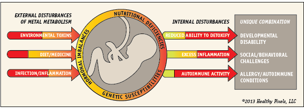
Heavy metal disturbance
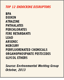
Some of the most promising areas of research reveal potential environmental factors of developmental disability. Exposure to mercury, cadmium, nickel, trichloroethylene, and vinyl chloride have been linked to autistic genetic mutations even before conception. Hundreds of chemicals and organic pollutants have been identified as endocrine disruptors, and they are very different in their composition and place in our environment. A list of the top endocrine disruptors is available through the Environmental Working Group.
Metals are among the many natural and synthetic endocrine disruptors that interfere with hormones as well as DNA methylation. Exposure to heavy metals has been associated with autism as well as other neurodegenerative diseases like Alzheimer’s and Parkinson’s diseases. The role of metals in autism relates to metal metabolism, one of the most intriguing and mysterious balancing acts in the body. The brain requires metals like zinc, iron, and copper to biologically function, while other metals like mercury, aluminum, cadmium, manganese and lead can accumulate and disrupt our internal environment. Both mercury and cadmium were the most consistently significant variables related to autism in one study. Deficiencies of zinc and magnesium were found in autistic infants.
Even minute quantities of metal, often called trace elements, can accumulate and interfere with many different organs, skin, and hormones in very different ways, making it difficult to trace the initial cause. For example, mercury exposure associates with reduced thyroid hormones, while cadmium associates with elevated thyroid hormones.
Internal interference
We cannot utilize heavier metals like aluminum, lead, and mercury, and our body must remove them through internal processes called methylation and transsulfuration.
“Methylation and transsulfuration play a crucial role in the synthesis of neurotransmitters, the generation of creatine, cell differentiation, embryonic development and the genesis of cell membranes. Moreover, disruption of methylation biochemistry can have a profound effect on epigenetics.” – North American Journal of Medicine and Science, 2012
Metals interfere with methylation and transsulferation in multiple ways, creating oxidative stress that taxes the body’s natural antioxidants for detoxification. One of these important products is glutathione, formed in the body from L-cysteine, L-glutamate, and glycine. Glutathione supports other antioxidants like vitamins C and E as well as iron. The immune system, nervous system, and gastrointestinal system are dependent on glutathione to function properly, and the brain requires it for protection.
Autistic types are “hypomethylators,” demonstrating a reduced ability to methylate and detoxify harmful substances. Most likely, their genetic differences determine the level of impact of metals such as mercury and selenium on glutathione-related activity. Glutathione is significantly reduced in autistic brains, allowing for increased oxidative stress, chronic inflammation, and DNA damage. Furthermore, oxidized glutathione is increased in autism, suggesting that autistic brain tissue is unusually burdened with toxins and lacking normal protection. In this manner, oxidation from environmental forces may create a cycle of brain inflammation that may aggravate the autistic condition.
DNA methylation is also the “copy machine” process that ensures our genes are properly replicated, and it is altered in the presence of heavy metals and industrial chemicals. In this manner, it appears that metals change our genetic programming in ways that favor disease.
Environmental exposure
Determining our dietary exposure to metals is enormously complicated, especially when food and vaccine labels lack complete information about sources, trace minerals, and safety. Even the Total Diet Study, routinely conducted by the FDA, analyzes only a small selection of foods and does not test for aluminum, chlorine, or fluoride. Furthermore, the EPA does not require compliance for levels of aluminum, manganese, chloride, fluoride, and other potential neurotoxins in our drinking water. According to the USGS scientific investigation report on domestic well water, “concentrations of aluminum, iron, and manganese were greater than USEPA SMCLs (secondard maximum contaminant levels) in about 7 to 21 percent of wells.” Arsenic, boron, manganese, strontium, and uranium also exceeded health benchmarks in about 1 to 7 percent of sampled wells. At higher levels, some of these trace elements can influence child behavior.
Accumulating research also points to the impact of pollution on autism, particularly through maternal and early life exposure to traffic congestion. A closer investigation revealed that perinatal exposure to diesel and mercury pollutants were associated with autism in both boys and girls. Another study showed risk with maternal exposure to disinfectants as well as exhaust and combustion products. It is notable that diesel, gasoline and lubrication oils are hazardous sources of metal oxide particles that may affect health.
Parental occupation has not been well studied, though exposure to asphalt and solvents show an association with autism. A larger study confirmed a clear association between ADHD behavior and prenatal exposure to solvents.
Malicious mercury
One of the most controversial metals surrounding autism is mercury. Some types of mercury accumulate in the body more than others, working with other toxins to possibly cause damage related to autism. The form of exposure such as inhalation, food consumption, and injection can also dramatically affect the amount of accumulation. Blood levels of mercury are generally higher in individuals diagnosed with autism; in hair samples of autistic children, mercury concentrations were significantly correlated with symptom severity.
There are numerous environmental sources of mercury that could potentially combine with other elements and pose a risk the body and brain. It is important to note that not all mercury is created equal. Mercury has very different health effects on the body depending on the compound, form of exposure, individual susceptibility, and environment!
Metallic or elemental mercury was once used in thermometers and potentially toxic when evaporated and inhaled. Mercury was used extensively in gold mining, and residues from the Gold Rush now pose a threat to the fish and water quality in several areas of California.
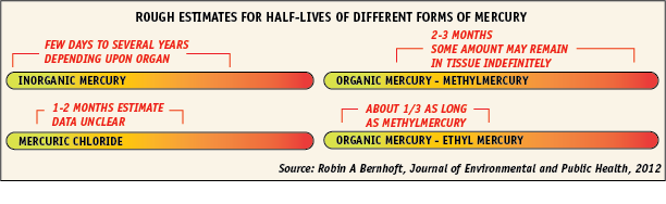
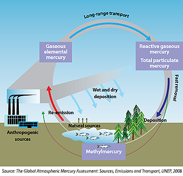
Inorganic mercury is a combination of mercury and other elements such as chlorine, sulfur, or oxygen. Mercuric chloride is an inorganic mercury compound associated with testicular damage resulting from oxidative stress. Inorganic mercury is released into the atmosphere from industrial sources such as coal burning plants or natural sources such as volcanic eruptions. Even cement manufacturing, waste management, cremation, and dentistry add mercury to our environment. Mercury particles may travel and be deposited locally or across very long distances – even between continents – before depositing on land or ocean through rain and snow, particularly in the Arctic and eastern United States. Mercury is also four-times higher in lakes near major US cities, demonstrating the serious impact of urbanization and power plants on the local ecosystems. Mercury deposits in soils and vegetation and then moves into adjacent aquatic environments or re-emits into the atmosphere by water and soil erosion. In these environments, mercury combines with carbon and becomes a more toxic organic mercury.
There are many different forms of organic mercury which show higher retention in the brain than inorganic forms. The most controversial form of organic mercury surrounding the autism-vaccine debate is ethyl mercury, a preserving, antibacterial ingredient commonly named thimerosal in certain flu vaccines in the US and a variety of others used before 2001. Mercury content of the flu vaccines is typically 25 micrograms, and oddly the EPA has no toxicity reference value for this type of mercury. Ethyl mercury can be found in various nasal sprays, antibiotics for the eye, ear preparations, and mascara.
The notoriously toxic methylmercury requires an even longer half-life than ethyl mercury, though some may theoretically remain in certain tissue indefinitely. Methylmercury bioaccumulates in living tissue, reaching hazardous levels in various organisms such as certain fish and shellfish. Both ADHD and autism appear to have strong associations with prenatal methylmercury exposure.
 The mystery of methylation
While directly consuming methylmercury through fish is known to be a health hazard, we aren’t quite sure if other forms of mercury can convert to methylmercury in the body. In fact, we aren’t even sure if the half-life of mercury can provide a safe measuring stick, as it does not indicate the effect on tissue or immune programming. In order to better understand what’s inside, we can start to look outside for clues.
In soil, sulfur chemistry is one of the strongest factors in the transformation of inorganic mercury to the more toxic methylmercury. Bacteria is the critical catalyst in this conversion process, with new types being discovered in rice paddies, anaerobic wastewater treatment plants, peat bogs, and even the body. Sulfate-reducing bacteria is the most common methylmercury generator, one of the oldest microorganisms on the planet. This familiar compound that smells like rotten eggs can bind with other compounds and metals to render them less toxic to living organisms.
Interestingly, autism associates with reduced sulfate in the body, though it has not been established if this relates to a cause or effect of environmental toxins. Furthermore, sulfur-reducing bacteria populate the gut in about half of the human population. Sulfate-reducing bacteria rely on sulfate for energy and release the waste product as hydrogen sulfide. Hydrogen sulfide, both toxic and beneficial to the body, may also play an important role in metal detoxification.
It is alarming to think that our gut bacteria and fungus might contribute to our vulnerability to methylmercury accumulation. While minute amounts of inorganic mercury are typically released rather quickly by the body, high levels of fungus (yeast) and bacteria like staphylococci, streptococci, and E. coli can actually turn mercuric chloride into the more toxic methylmercury. Interestingly, other studies could not find a similar conversion of cinnabar (mercuric sulfide) to methylmercury with gut bacteria, indicating the importance of researching the specific forms of mercury responsible for toxic transformations inside the body.
While it is outside the scope of this report, it is worth considering the regions of the world where methylmercury readily accumulates in the food chain. Wetlands, newly flooded regions, small warm lakes, and areas of the Everglades exposed to sulfur contamination are particularly vulnerable to methylmercury accumulation. Streams that don’t drain into wetlands are also particularly high in methylmercury due to bacterial activity, warm temperatures, and phosphorus removal. Recent studies have even found methylmercury in coastal fog which deposits mercury on land and vegetation near the coast. We do not know if atmospheric or soil concentrations of methylmercury can affect the body, though consumption of contaminated fish in these areas may be an indirect consequence.
Sources of mercury pollution
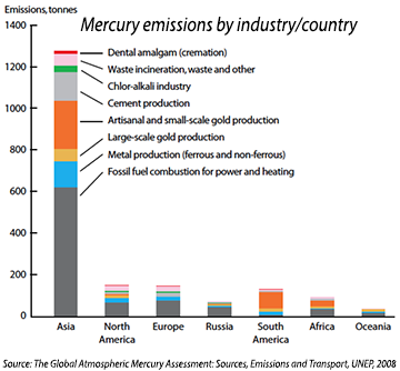
Power plants fired with fossil fuels are the primary source of manmade mercury emissions, though shifting environmental conditions and other human variables make it difficult to study their impact. Even so, researchers found that autism and special education rates in Texas were strongly correlated with local, environmentally released mercury. Other studies indicate that the timing of environmental exposure may be another factor.
“We discovered a trend where an increase in mercury was strongly related to an increase in autism prevalence three years later.” – 2009, Ninth IEEE International Conference on Bioinformatics and Bioengineering
Mercury can often travel around the globe in the upper atmosphere before depositing primarily through rainfall, snow, or fog and accumulating in ecosystems. It is surprising that the majority of mercury in the atmosphere is emitted by volcanic eruptions. However, living organisms are showing sensitivity to additional loads of this enduring element. In a biogeochemical model, scientists concluded that only 17% of the mercury in our surface ocean is from natural sources, and 50% of the manmade mercury originates from pre-1950 emissions.
“The accumulated burden of legacy anthropogenic Hg means that future deposition will increase even if primary anthropogenic emissions are held constant.” – Global Biogeochemical Cycles, 2013
Mercury by mouth
Fish: Larger, older fish including shark, tuna, swordfish, mackerel and halibut accumulate the most toxic form of mercury, methylmercury, but levels vary widely based on the levels in their prey and local water chemistry. Even smaller fish may be highly contaminated in lakes and streams based on their heavy metal content. The widespread concern over fish consumption is complicated by the lack of awareness, minimal mercury data, and large differences of toxicity among types of fish.
“Children are especially vulnerable and may be exposed directly by eating contaminated fish. Methylmercury bioaccumulated in fish and consumed by pregnant women may lead to neurodevelopmental problems in the developing fetus. Transplacental exposure is the most dangerous, as the fetal brain is very sensitive. Neurological symptoms include mental retardation, seizures, vision and hearing loss, delayed development, language disorders and memory loss.” – World Health Organization
Methylmercury can also be absorbed by lymph which poses serious threats to our immune system, including autoimmunity. Certain metals such as mercury appear to actually induce autoimmunity or aggravate autoimmune disease in genetically susceptible animals. Even gold and silver may produce autoantibodies, though the immune response is much weaker than mercury.
Breastfeeding cannot protect a baby from these toxins, according to animal studies where methylmercury in mother’s milk is nearly all absorbed and retained with very slow elimination and extremely high brain tissue concentration. However, certain nutritional aspects of uncontaminated fish may provide some protection. In one study, prenatal exposure to mercury was associated with greater risk of ADHD symptoms – particularly in boys – while fish consumption showed some protection. While there is evidence that prenatal mercury exposure affects brain and behavior, we have only scratched the surface of the problem.
Rice: Mercury from rice grown in mercury-polluted areas can also contain excessive levels of methylmercury. Flooded, wetland paddy fields favor anaerobic bacteria growth and acidic soil that may support methylmercury growth more than other crops.
High fructose corn syrup: Some researchers believe that the mercury from HFCS leads to altered genes, mineral imbalance, and increased inflammation that can be passed down through generations. High fructose corn syrup (HFCS) is used in a wide range of foods and beverages, and it is often manufactured with mercury cell chlor-alkali products to increase shelf life. As HFCS manufacturing is proprietary, it is impossible to locate the source of the mercury. One study found mercury levels up to .570 micrograms per gram of HFCS. With the US average daily consumption HFCS reaching 50g, the average daily mercury exposure from HFCS could potentially total 28.4 micrograms. This is well over the FDA’s acceptable daily intake for mercury at .4 micrograms per kg (about 18 micrograms for a 100 lb child).
Furthermore, high fructose consumption is highest in adults between during the early reproductive years. In an interesting study by the Institute for Agriculture and Trade Policy, mercury was detected in 60% of dairy samples – most chocolate milk – and 40% of dressings and condiments. Many chocolate syrups, dressings, and condiments such as ketchup contain high fructose corn syrup.
Dental fillings: Dental amalgams incorporate inorganic mercury, not methylmercury, and have been used extensively for the past 100 years without a direct link to autism. However, there is a negative impact on the thymus and its hormones among dental staff, and studies have started to examine fetal exposure to mercury. Researchers found that mothers receiving six or more amalgam fillings during pregnancy were three times more likely to have children diagnosed with severe autism than mild autism. When looking at amalgams, over 80% of Canadians are exposed to a daily dose of mercury that exceeds the Canadian reference exposure level, and millions more exceed EPA levels in the US.
Mercury by injection
Thimerosal, also known as ethylmercury, is an organic mercury compound used as an adjuvant – an additive in drugs, vaccines, and tattoo inks to kill bacteria and fungus. Thimerosal was widely used in Hep B vaccines between 1991-2001, and by 2005, all infants became routinely immunized at birth. Thimerosal was also used in formulations given to Rh-negative mothers within 72 hours of birth. Since the early 2000s, mercury-based thimerosal has been removed in most vaccines excluding the flu shot. The FDA maintains a current list of thimerosal-containing vaccines and their history.
While there is no direct correlation between thimerosal and autism, a very critical study found a link to tic disorders in boys. Studies also examined how thimerosal could irreversibly affect the brain’s production of serotonin and dopamine. In animal studies, thimerosal caused an excess of certain amino acids – glutamate and aspartate – along with a decrease of glycine and alanine.
Thimerosal has been a popular suspect in the autism mystery, but evidence of long-term effects is limited. The short-term effects and form of exposure may tell a different story. Thimerosal exposure in early infancy was linked to deficiencies in psychomotor development in the 12th and 24th month of life, though not longer. Additionally, animal studies showed that oral consumption of methylmercury resulted in far greater brain tissue accumulation than injections of thimerosal.
Foiled by aluminum?
While mercury has diverted our attention, it is worth discussing another common metal that is often overlooked. Aluminum is the most abundant metal on Earth, but it accumulates in the brain and has been linked to Parkinson’s and Alzheimer’s disease. Aluminum, a non-essential element, competes with essential minerals like magnesium, iron, and calcium, and wreaking havoc on our metabolism, immune system and brain function. New research described how aluminum may even contribute to inflammatory, autoimmune conditions like Crohn’s disease. Metals such as aluminum can also interfere with dopamine neuron degeneration – as evident in Parkinson’s disease. Dopamine and serotonin are two of the many neurotransmitters that are dysregulated in autism.
Unlike mercury, aluminum sticks around much longer than a few months. One study suggested that the half-life of aluminum in the brain may be around 12 years, and another study estimated 10-20 years. Like other metals, scientists have no way to measure accumulation and toxicity in live human brain tissue. Tests are limited to hair, blood and urine which do not accurately reflect the impact of metals on individual organs, bones and tissues.
Vaccines
“Experimental research…clearly shows that aluminum adjuvants have a potential to induce serious immunological disorders in humans. In particular, aluminum in adjuvant form carries a risk for autoimmunity, long-term brain inflammation and associated neurological complications and may thus have profound and widespread adverse health consequences. ” – Current Medicinal Chemistry, 2011
Aluminum salts have been used as adjuvants in vaccines for nearly a century, stimulating the immune response in ways that scientists still cannot explain. Aluminum somehow boosts the efficacy and strength of our vaccines, but it may also contribute to autoimmune disease later in life. Aluminum disrupts iron metabolism in the intestines and can lead to anemia, inflammation, and obesity. Recent studies revealed that aluminum causes cells to die and release their DNA. If a body has elevated autoantibodies against parts of these dying cells, the immune balance can shift to become pro-inflammatory. A body can develop autoantibodies to almost anything, defining the target as an allergen and responding in a subtle or not-so-subtle manner. The number of variables in this cellular process is enormously complex and has only recently been examined as the process of NETosis (neutrophil extracellular trap).
Alarmingly, babies born today receive .25 mg of aluminum in their Hep B vaccine given on the day of birth. Kids are getting more than four times the number of vaccines than they received in the 1970s, spiking aluminum levels as early as 6 weeks of age (as in the case of Pediarix) with combination injections for convenience. The rise in autism cases has strongly correlated with recommended childhood vaccinations since 2001.
A close investigation showed that: (i) children from countries with the highest autism spectrum disorder (ASD) prevalence appear to have the highest exposure to Al from vaccines; (ii) the increase in exposure to Al adjuvants significantly correlates with the increase in ASD prevalence in the United States observed over the last two decades (Pearson r = 0.92, p < 0.0001); and (iii) a significant correlation exists between the amounts of Al administered to preschool children and the current prevalence of ASD in seven Western countries, particularly at 3–4 months of age (Pearson r = 0.89–0.94, p = 0.0018–0.0248). – Journal of Inorganic Biochemistry, 2011
Studies on primates showed that vaccines limited development in certain parts of the brain, impacting motor neurons cells that control body movements and spatial memory. Serious conditions like Gulf war syndrome have been linked to exposure to aluminum adjuvants, leading scientists to propose the new classification ASIA – Autoimmune Syndrome Induced by Adjuvants.
Foods and products
Aluminum can also be absorbed in the gut or skin from certain baked goods, processed cheese, frozen foods, baby clams, fish such as canned tuna, tea, buffered aspirin and antacids, cookware, deodorant, baby wipes, cosmetics, and lotions. Most of us don’t recognize the many names for aluminum compounds such as alum – a potassium aluminum sulfate salt used in “natural” deodorants and pickling. Food colorings that list “aluminum lake” are composed of aluminum hydroxide and are widespread, appearing in the popular “Flintstones” kids vitamins and “M&Ms” candy. Cosmetics and pharmaceuticals use many different clay minerals that contain aluminum.
Surprisingly, aluminum is an approved additive and can occur at levels of 180 mg/serving in some foods! The Agency for Toxic Substances and Disease Registry determines the intermediate-duration oral minimal risk level (MRL) at 1 mg Al/kg/day – this converts to only 45 mg of Al for a 100-lb person in one day. Aluminum levels were high in tested products including frozen pizza, non-dairy creamer, baking powder, pancake/waffle mixes, and ready-to-eat pancakes. Aluminum cookware can dramatically raise levels in food as a result of temperature, acidity, salt content, and length of cooking.
Scientists have recognized the leaching of aluminum from containers and cookware for almost a century. In 1996, aluminum was discovered in beverages stored in glass bottles, while levels in beverages from steel cans and aluminum cans were even higher. Glass contains minute quantities of aluminum and other metals bound in the silicon, and heat-sterilization can speed up the leaching process. This can be demonstrated in studies of sterilized cow’s milk which contains higher levels of aluminum than raw milk. Recent analyses found that the amount of aluminum increases with decreasing levels of formula concentration, so ready-to-use liquid formulas have more aluminum content than powders – presumably from manufacturing processes and increased contact with the container. Glass storage containers appear to leach more aluminum than steel containers.
Intravenous feeding or parenteral nutrition utilizes glass containers for storing solutions, and parenteral nutrition is commonly used for infants born prematurely or with low birth weight.
“Parenteral nutrition (PN) has long been implicated as a major source of aluminum exposure due to contamination of the component ingredients. PN component products are contaminated with aluminum in raw materials as well as byproducts from the manufacturing process, where aluminum leaches from glass vials during autoclaving.” – Nutrients, 2012
In fact, the aluminum levels in PN do not meet the FDA “safe limit” of less than 5μg/kg/day! The aluminum contamination of parenteral nutrition solutions appears largely related to nutrients calcium gluconate, inorganic phosphates, and cysteine hydrochloride. Other studies noted that that aluminum content was correlated with calcium in the solutions.
Infant formula
Aluminum exposure can be very high in infant formulas, particularly soy-based powdered forms that can result in consumption of over 100 mg of aluminum per year! About 36% of infants in the US receive soy-based infant formula in the first year of life. Though soy formula was first introduced in 1929, formula was not heavily marketed to the public until 1988 amidst criticism from the American Academy of Pediatrics. While studies have not examined aluminum in autism, the condition was determined significantly more likely in children who were not breastfed.
Even vitamins and infant formula can be an unsuspecting source of unpredictable heavy metal chemistry that affect body and brain processes. Even mineral compounds including iron and copper can stimulate the oxidation of antioxidant organic compounds like vitamin E. An interesting study linked the introduction of advertised infant formula in 1988 to the subsequent rapid increase of autism. Exposure to excess multivitamins can lead to neurotoxicity and metabolic imbalances in vulnerable children, and we desperately need to better determine optimal amounts of each vitamin for both the fetus and growing child.
Infant formula is made with soy protein created from highly processed soybeans that may be high in aluminum for a variety of reasons. Soybeans naturally contain a high amount of aluminum; certain additives such as calcium and phosphorus somehow increase levels; metal processing equipment can taint the formula; and glass containers can leach aluminum with heat-treatment. The manufacturing method can include an alcohol wash, acid wash, or heat-treatment which can involve acid tanks.
A recent in-depth evaluation of soy infant formula by the National Toxicology Program overlooked both cognitive effects and aluminum toxicity entirely. The expert panel was searching for toxicity from the isoflavones, genistein, daidzein, and glycitein – not aluminum or manganese. Consequently, there was no mention of potential toxicity from aluminum in their 789-page report. Regarding brain development, the report states “Two cognitive function studies were reviewed for this report and were considered of no utility.” Alternatively, the European Society for Paediatric Gastroenterology Hepatology and Nutrition cautions that soy-based formula has nutritional disadvantages and contains “high concentrations of phytate, aluminum, and phytoestrogens, the long-term effects of which are unknown.”
Aluminum compounds
Aluminum appears to increase toxicity when interacting with a variety of substances. Aluminum combines with fluoride in the body and can associate with symptoms of autism and immune dysregulation. Infants can be exposed to fluoride in drinking water, toothpaste, and even fluoride drops which are routinely prescribed to prevent cavities. Other substances such as malate (malic acid is high in apples, nectarines, grapes and wines, gum, and sour candy) and citrate (citric acid is high in soft drinks, vitamins, and citrus fruits) may affect toxicity by enhancing aluminum absorption in the gut. As indicated in animal studies, glutamate in the diet (including MSG or high-glutamic acid foods like seaweed, soy sauce, cheese) may increase aluminum absorption in the brain.
Aluminum exposure may be most frequently experienced through aluminum sulfate salts for purifying drinking water and treating wastewater. We desperately need to research the influence of aluminum compounds in the womb given our dependency and constant exposure.
Lead exposure
Lead exposure inhibits energy metabolism and contributes to neurological dysfunction. Cases of ADHD have been strongly associated with lead exposure, even in very low amounts. Reports describe autistic-like symptoms in children with lead poisoning, and genetic studies find that autistic children process the heavy metal differently than others. Higher levels of lead and other toxic metals were found in blood and urine. Children are particularly vulnerable to lead as they explore a variety of surfaces with their hands and mouths. Most exposure to lead comes from dust, particles or even soil contaminated with lead paint used prior to 1978. Old plumbing used in the 1930s may have contained lead that slowly leaches into the water. Even new pipes may be joined with lead-based solder that contaminates the drinking water water. Imported toys can also contain lead. Broth made of animal bones which accumulate lead and other metals has been associated with exposure.
As with other toxins, autistic individuals metabolize lead differently and this appears controlled by genes that regulate inflammation and immune health. It is not yet clear if lead is a factor in ADHD and autism without genetic susceptibility.
Manganese toxicity
Though manganese is essential in our diet for metabolism and bone growth, high levels are associated with motor and behavioral problems in children. Parenteral nutrition (IV feeding) can also introduce high levels of manganese. High levels are associated with a Parkinson’s-like disorder in adults and learning and behavioral challenges in children. Studies found that higher levels of manganese in drinking water, not diet, was correlated with lower IQ scores in children. A large study of children in Bangladesh correlated both manganese and arsenic in drinking water with reduced intellectual function.
Sources of manganese exposure can include soy-based infant formula, air pollution, fungicide residue, and drinking water. Researchers found that daily consumption of rice could easily expose someone to levels of manganese and zinc that exceed the RDA. Shower exposure to water high in manganese may also increase neurotoxic risk. Soy-based formula can contain 100 times the amount of manganese as breast milk. Even soybean yogurt showed much higher concentrations of manganese, copper and nickel compared to dairy yogurt. Researchers have demonstrated that manganese from soy-based infant formula can be excessively absorbed into the brain, potentially impairing development. One very elaborate study of manganese absorption noted that suckling infants appeared to accumulate more manganese from inhalation than diet. Interestingly, the psychostimulant methylphenidate used to treat ADHD also appears to lower manganese levels and increase dopamine.
Cadmium concerns
In one recent study, children with high levels of urinary cadmium expressed more learning disabilities and need for special education. According to WHO, high levels of cadmium can be found in certain oysters, scallops, mussels, and crustaceans. Cadmium, lead, and other heavy metal consumption can result from exposure tobacco smoke, contaminated water from mining areas, and even rice crops in contaminated soil. Like other metals, cadmium can be transported in the atmosphere and concentrations in our air and soil can differ greatly between territories.
Other disruptive toxins
Not all studies have been able to clearly implicate metals to autism and other developmental disorders. Other endocrine disruptors may also influence susceptibilities and conditions, particularly thyroid hormone function and GABA mechanisms.
“…lead, methylmercury, pesticides, tobacco (cotinine), persistent organic pollutants such as PCBs, and environmental hormones such as bisphenol A and phthalates have … indicated association between neuronal disability and exposure levels in children.” – National Institute for Environmental Studies, Japan, 2012
Pesticides
Exposure to certain pesticides is being linked to higher risk of autism. Other studies link ADHD with early exposure to organophosphate pesticides – even in the womb. Pregnant mothers living within 500 meters (1/3 mile) of fields treated with organochlorine agricultural pesticides showed an increased risk for have autistic children. Pesticides are not limited to crops; mothers of autistic children were twice as likely to have used certain pet shampoos during their child’s early life.
Diet is the source of most pesticide exposure in children, with wheat and corn being the top two most likely sources of organophosphates. Experimental evidence shows that pesticides, like metals, may alter DNA methylation to increase disease risk. A list of pesticides that alter genetics or disrupt DNA methylation includes certain endocrine disruptors, persistent organic pollutants, metals, herbicides, insecticides, and fungicides. In addition, organochlorine pesticides were strongly linked to lower vitamin D levels. More research is needed to understand how pesticides in the air, water and food can contribute to diseases of the brain and body.
Phthlates
Phthalates are used as plasticizers and solvents in tubing, personal care products, and pesticides. Recent studies have identified a link between urinary phthlate concentration and autism. Prenatal exposure to phthlates was associated with ADHD-like behavioral problems in young children.
Contemplating cause or effect
Currently, perinatal endocrine disruptors appear to be the primary suspects for genetic and hormone disturbances that may increase susceptibility to autism and ADHD. “Perfect embryonic storms” could theoretically include a combination of heavy metals, industrial chemicals, and organic pollutants that results in the incredible diversity of symptoms found in autism, ADHD, and other behavioral conditions. This complexity demands a much more refined approach to study. As researchers Georgiades, Szatmari and Boyle propose:
“…heterogeneity could provide a general framework that will guide the development, implementation and interpretation of new study designs and measurements and that will have the ability to capture individual and subgroup differences within autism.” – Neuropsychiatry, 2013
***
The next report in this multi-part series will propose prevention guidelines that consider current research on environmental toxins and nutritional factors.

Leave a Reply